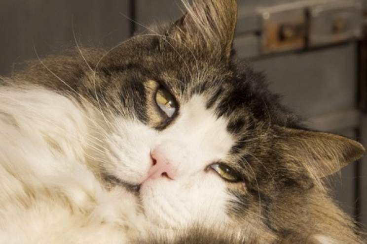
Cat owners will certainly know what we are talking about. The third eyelid is the whitish structure that sometimes seems to emerge from the inner corner of the eye, covering a greater or lesser part of it. But what is it for, and why is it sometimes visible and sometimes not?
What Is the Third Eyelid?
Third eyelid is the common name used to refer to the nictitating membrane. It is effectively an additional eyelid alongside the upper and lower eyelids. It begins at the inner corner of the eye, near the nose, and extends towards the outer side.
Its presence is particularly significant in reptiles and birds, which have a highly developed third eyelid capable of completely covering the eye when necessary. Dogs, and especially cats, also have a fairly large third eyelid, which can cover up to half of the eye’s total surface, but is nevertheless opened only on a few occasions. Humans have one too, but it is so underdeveloped as to be almost irrelevant.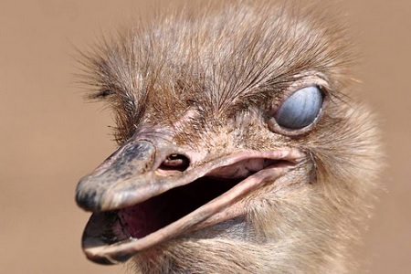
The main function of the third eyelid is to protect the eye, which is a very delicate organ. In particular, reptiles open it completely when they want to keep the eye moist and protected while still maintaining good visibility of their surroundings. Put simply, it is as though animals could wear contact lenses to prevent sand, dust and foreign bodies from scratching the eye and to prevent tears from evaporating, causing troublesome problems. This structure also helps produce some of the tears, thanks to the presence of special glands.
The third eyelid is easy to recognise, especially in cats. If it is not affected by any direct problem, it appears as a shiny, whitish membrane that is rather wrinkled and moves horizontally, covering the inner corner of the eye.
Dogs and cats normally “open” the third eyelid only when they are sleeping. In fact, if we observe our friend while deeply asleep, they may keep their eyes slightly open, especially if they are dreaming. In these cases, we may have the impression that the eye has turned white, but it is actually the third eyelid covering the visible portion of the eye.
However, a dog or cat may expose it while awake, or the membrane may even acquire particular characteristics as a result of certain conditions or diseases.
Why Does the Third Eyelid Become Visible?
When the third eyelid becomes visible, this is called protrusion. The membrane looks normal, but remains visible in one or both eyes even when the animal is awake. The main reason, especially in cats, is linked to pain. Felines experiencing pain anywhere in the body tend to “open” the third eyelid.
In cats, this may also mainly involve Haw’s syndrome, which affects both eyes and may be caused by:
-
Intestinal parasites, especially tapeworms
-
Eye inflammation
-
Eye trauma
-
Neurological problems
-
Infections
-
Gastrointestinal problems
The third eyelid may instead look different from usual. The most common changes include:
-
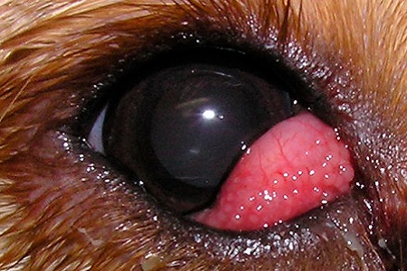
“Cherry eye”: this term refers to eversion or prolapse of the third eyelid, specifically the tear gland contained within it. This gland is normally hidden, but in some cases it may protrude and become visible externally. In these cases, we will notice a prominent, swollen red structure, which gives the condition its name. It is more common in dogs, particularly in certain breeds such as Bulldogs, Beagles, Cocker Spaniels and other flat-faced dogs. The causes are unknown, but it is thought to result from severe inflammation or irritation of the gland, as well as genetic factors linked to the breed;
-
Eversion of the third eyelid: in this case, the membrane literally turns over, folding outward. It may occur particularly in Burmese cats and large-breed dogs, especially during the first year of life. It usually disappears as the animal grows, but in some cases surgical removal may be required;
-
Conjunctivitis of the third eyelid: the membrane develops severe inflammation, mainly due to bacteria or allergies, and therefore appears swollen and reddened;
-
Injuries: the third eyelid, like the other structures of the eye, may be subject to trauma and injury, for example from scratches or foreign bodies;
-
Tumours: the third eyelid may also be affected by tumours, both benign and malignant.
What to Do
In these cases, a veterinary examination is essential. The veterinarian can perform a series of assessments and investigations to identify the cause of the problem. Once a diagnosis has been reached, treatment will vary according to the type of condition or disease.
In general, if the membrane shows no visible changes, a stool examination may be necessary to check for intestinal parasites, for which the animal will need to be dewormed. Neurological or blood tests may then be performed to determine whether the animal has injuries or problems that could be causing pain.
When the third eyelid shows changes, such as cherry eye or eversion, it may be necessary to use targeted treatments, including antibiotics or anti-inflammatory drugs, or even surgery to reposition it or remove it partially or completely.
Some diseases of the third eyelid can cause the animal considerable discomfort, but above all they may expose the eye to a series of serious complications, so never neglect the problem.